Understanding Dental X-ray Radiation: Safety, Benefits, & Myths

In the realm of modern dentistry, dental x-ray radiation has become an indispensable tool for accurate diagnosis and effective treatment planning. While concerns around radiation exposure are common, advancements in technology and strict safety protocols have made dental radiography safer than ever before. This comprehensive guide aims to provide detailed insights into dental x-ray radiation, its benefits, potential risks, and the revolutionary methods used to minimize exposure, ensuring that patients and practitioners can make informed decisions about dental imaging procedures.
What Is Dental X-ray Radiation?
Dental x-ray radiation refers to the ionizing radiation emitted during radiographic imaging of the teeth, gums, and jawbones. These images provide vital information that cannot be obtained through visual examination alone, such as hidden tooth decay, bone loss, impacts of trauma, and other dental conditions.
The radiation used in dental x-rays is a form of electromagnetic energy, similar to visible light but with much higher energy levels capable of passing through tissues to produce images on photographic film or digital sensors. Modern dental clinics utilize state-of-the-art equipment designed to minimize radiation exposure while maximizing image quality.
The Science Behind Dental X-ray Radiation
Ionizing radiation used in dental x-rays works by ionizing atoms in tissues, which creates images based on variations in tissue density. Dense structures such as enamel and bone absorb more radiation, appearing lighter on images, while soft tissues and areas with less density appear darker.
- Digital X-ray Technology: Uses electronic sensors instead of traditional film, reducing radiation doses significantly.
- Film X-ray Technology: Generates composite images, but with higher doses compared to digital methods.
- Advanced Equipment & Techniques: Include rectangular collimators, faster film speeds, and protective shields implemented to reduce radiation.
Safety Measures and Protocols in Dental Radiography
Quality dental practices prioritize patient safety through comprehensive safety protocols. These measures ensure that radiation exposure remains as low as reasonably achievable (ALARA principle). Some key safety measures include:
- Use of Protective Aprons and Thyroid Collars: Shields vital organs from scatter radiation during the imaging process.
- Digital Imaging: Significantly reduces radiation doses compared to traditional film x-rays.
- Precise Technique and Equipment Calibration: Ensures optimal image quality with minimal exposure.
- Limiting Exposure Frequency: Avoiding unnecessary recurrent imaging, especially in vulnerable populations like children and pregnant women.
- Proper Training for Practitioners: Ensures technicians are skilled in minimizing radiation exposure while achieving diagnostic quality images.
Common Types of Dental X-ray Images and Uses
Understanding the different types of dental x-rays helps in grasping their importance and safety considerations:
- Bitewing X-rays: Show the crown of upper and lower teeth simultaneously, excellent for detecting cavities between teeth and monitoring bone levels.
- Periapical X-rays: Capture the entire tooth, from crown to root tips, useful for diagnosing root and bone issues.
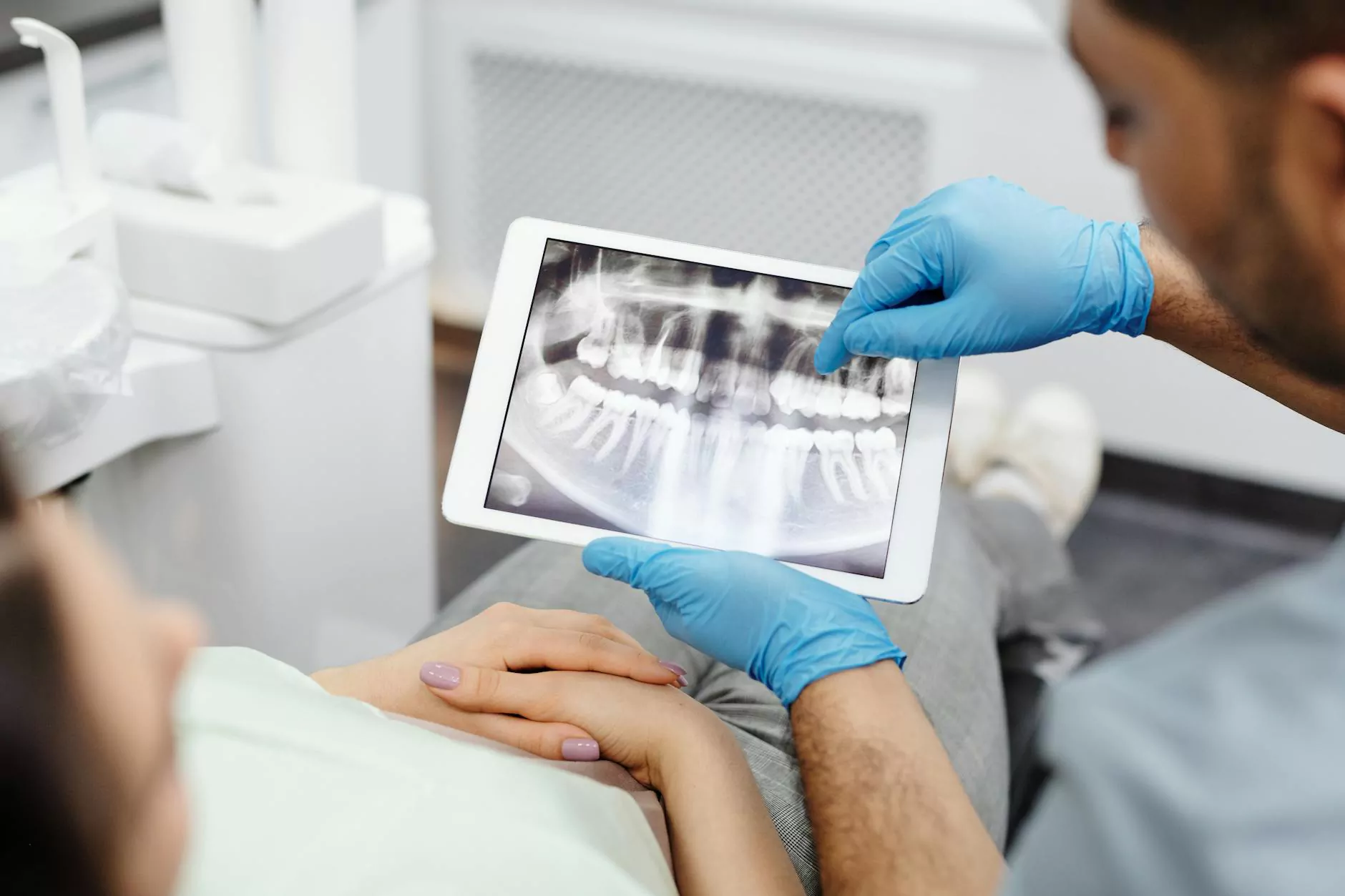
- Pano or Panoramic X-rays: Provide a broad view of the jaw, teeth, sinuses, and temporomandibular joints (TMJ), aiding in comprehensive treatment planning.
- Cephalometric X-rays: Mainly used in orthodontics to analyze the jaw and facial structure.
The Benefits of Dental X-ray Imaging
Despite misconceptions about radiation risks, the benefits of dental x-rays far outweigh potential harms when performed with proper safety protocols. They enable:
- Early detection of dental caries, preventing more extensive decay.
- Assessment of the health of the roots, jawbone, and surrounding tissues.
- Detection of abnormalities like cysts, tumors, and infections.
- Monitoring the progression of periodontal disease.
- Guidance for complex procedures such as implants and orthodontics.
Addressing Myths and Concerns About Dental X-ray Radiation
Many patients hold misconceptions about the safety of dental x-ray radiation. It's crucial to address these confidently to promote informed decisions:
Myth 1: Dental x-rays expose patients to dangerous levels of radiation
Fact: Modern dental x-rays involve very low doses of radiation. Digital imaging and safety protocols reduce exposure to levels comparable to, or even less than, natural background radiation encountered in daily life.
Myth 2: Pregnant women should avoid dental x-rays
Fact: Routine dental x-rays are safe during pregnancy when proper shielding and precautions are used. Dentists typically recommend postponing non-essential imaging until after pregnancy unless necessary for urgent diagnoses.
Myth 3: Repeated dentals x-rays cause cumulative health issues
Fact: When performed judiciously, with proper safety measures and limit on frequency, digital x-rays pose minimal cumulative risk.
Advances in Dental Imaging Technology and Reduced Radiation Exposure
Technological progress has continually enhanced the safety and efficacy of dental imaging:
- Digital Radiography: Cuts radiation dose by up to 90% compared to traditional films.
- High-Speed Films and Phosphor Plates: Increase image capture speed, reducing radiation needed.
- Collimation Techniques: Focus the x-ray beam precisely, limiting scatter radiation.
- 3D Imaging and Cone Beam CT (CBCT): Provide detailed 3D views with controlled radiation doses tailored to specific needs.
Guidelines for Safe Dental X-ray Use
Dental practices adhere to strict regulations and best practices to safeguard patients:
- Perform only when clinically justified.
- Use the minimal effective radiation dose.
- Employ shielding and protective equipment.
- Limit the frequency of imaging, especially in children and pregnant patients.
- Ensure practitioner training in radiologic safety.
Why Choose 92Dental.co.uk for Your Dental Imaging Needs
At 92Dental.co.uk, we prioritize patient safety while providing state-of-the-art dental imaging services. Our clinic employs the latest digital x-ray technology, ensuring:
- Minimal Radiation Exposure: Our equipment is calibrated for optimal safety and image clarity.
- Highly Trained Staff: Experts trained in radiologic safety protocols.
- Comfortable Environment: We ensure your experience is seamless, safe, and reassuring.
- Comprehensive Care: From routine check-ups to complex diagnostics, we tailor imaging to your needs.
Informed Decisions: The Role of Patients in Dental Imaging Safety
Patients should engage actively in understanding their dental imaging options. Key tips include:
- Discuss the necessity of x-rays with your dentist.
- Ask about the type of imaging and safety measures used.
- Inform your dentist if you are pregnant or have specific health concerns.
- Follow recommendations on frequency and safety precautions.
Conclusion: The Future of Safe Dental Imaging
As dental technology evolves, the focus remains on balancing diagnostic benefits with safety. Innovations like digital radiography and 3D imaging continue to reduce dental x-ray radiation exposure while enhancing diagnostic capabilities. Patients and practitioners are empowered by these advancements to make well-informed choices, ensuring dental health is maintained with the utmost safety.
Whether you need routine examinations, complex diagnostics, or preventive care, modern dental radiography offers unmatched precision with minimal risk. Trust in practices like those at 92Dental.co.uk, where safety, comfort, and excellence come together for your dental health journey.
dental x ray radiation